Image Integrity
Table of contents
Preparing images
The final image must correctly represent the original data and conform to communitystandards.
Images gathered at different times or from different samples should not be combined intoa single image.
If juxtaposing images is essential, the borders should be clearly demarcated in the figure, labelledand described in the legend.
Any elements added to images, such as insets, should be clearly indicated. The use of touch-up tools, such as cloning and healing tools in Photoshop, or any feature that deliberately obscures image data, is to be avoided. The use of any sort of generative AI in figures is not permitted. This includes content-aware editing tools in Photoshop.
Photoshop
Do not use these tools in Photoshop on image data:
- Spot Healing Brush Tool
- Remove Tool
- Healing Brush Tool
- Patch Tool
- Content-Aware Move Tool
- Eraser Tool
- Background Eraser Tool
- Magic Eraser Tool
- Clone Stamp Tool
- Generative Fill
Adjustments, such as changes to contrast, should be applied to the entire image, andapplied equally to controls.
Please check for image duplication within your figures. If a panel is intentionallyduplicated then it must be clearly labelled to indicate that.
If you notice any errors or duplicate images within your figures at any time, please let usknow so that the error can be corrected.

Gels
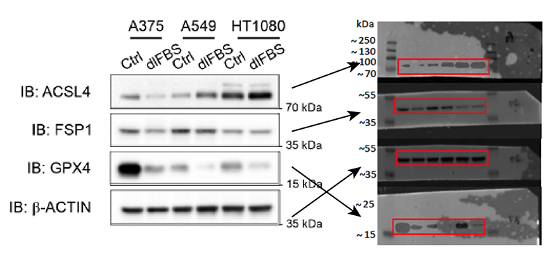
Please provide original uncropped data for all of the gels used in your final figures. Contrast, exposure, and dimensions in these should match the gels in your figure as closely as possible.
Please order the raw gels in the same way that they appear in the figure, and label each gel as clearly as possible.
Images of raw, uncropped gels should be provided that accurately represent the intensity and contrast of bands in the figure.
More than one raw gel image can be provided to show varying exposures, and to show how the image was processed.
Avoid: raw gels that are not clearly arranged and labelled and do not represent the gels in the figure accurately. 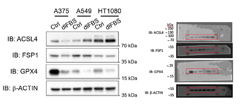
Recommended: clearly labelled raw gels arranged in the same order they appear in the figure.


If lanes of a gel are spliced together vertically this must be clearly marked with separation or a line.
Avoid: splicing of gels that is not clearly labelled. 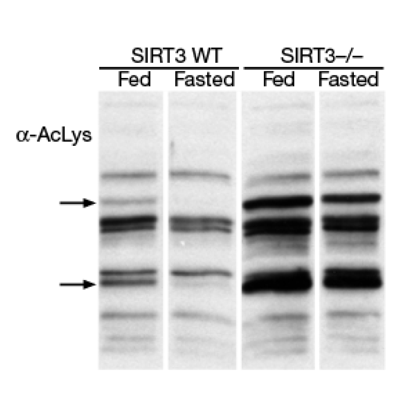
Recommended: white or black lines added between gels to indicate splicing.


Avoid horizontal splicing within a single gel panel; the parts should be separated and the weights clearly labelled.
Do not mask any elements within your gel or use any tools to remove or add data.
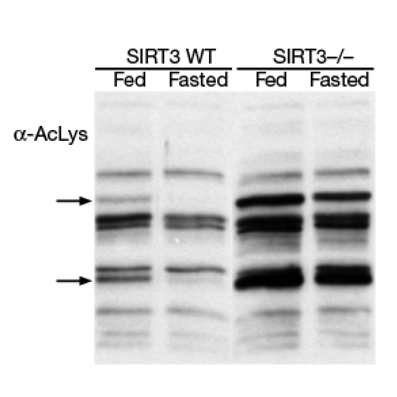
Do not adjust contrast in a way that removes any bands or information. Do not increase contrast on your gel images so that background details are lost.
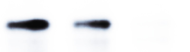
Avoid: adjusting contrast so that information is lost. 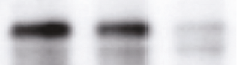
Recommended: present the image showing all data.


Do not adjust contrast to individual bands or parts of a gel in a way that alters their intensityrelative to other bands in the same gel.
Please note that your gels could be checked for any signs of improper manipulation or duplicationacross figures.